Ongoing research is being conducted to develop simple and inexpensive paper-based diagnostic tests for early cancer detection. These tests, also known as “paper-based biosensors,” employ a small strip of paper that changes color in response to specific cancer biomarkers or other molecules.
Engineers designed a nanoparticle sensor that could enable early diagnosis of cancer with a simple urine test. The sensors, which can detect many cancerous proteins, could also be used to distinguish the type of a tumor or how it is responding to treatment.
MIT engineers have designed a new nanoparticle sensor that could enable early diagnosis of cancer with a simple urine test. The sensors, which can detect many different cancerous proteins, could also be used to distinguish the type of a tumor or how it is responding to treatment.
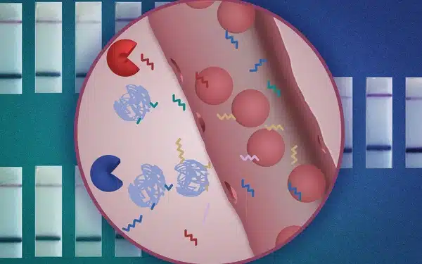
The nanoparticles are designed so that when they encounter a tumor, they shed short sequences of DNA that are excreted in the urine. Analyzing these DNA “barcodes” can reveal distinguishing features of a particular patient’s tumor. The researchers designed their test so that it can be performed using a strip of paper, similar to an at-home Covid test, which they hope could make it affordable and accessible to as many patients as possible.
We are trying to innovate in a context of making technology available to low- and middle-resource settings. Putting this diagnostic on paper is part of our goal of democratizing diagnostics and creating inexpensive technologies that can give you a fast answer at the point of care.
Sangeeta Bhatia
“We are trying to innovate in a context of making technology available to low- and middle-resource settings. Putting this diagnostic on paper is part of our goal of democratizing diagnostics and creating inexpensive technologies that can give you a fast answer at the point of care,” says Sangeeta Bhatia, the John and Dorothy Wilson Professor of Health Sciences and Technology and of Electrical Engineering and Computer Science at MIT and a member of MIT’s Koch Institute for Integrative Cancer Research and Institute for Medical Engineering and Science.
In tests in mice, the researchers showed that they could use the sensors to detect the activity of five different enzymes that are expressed in tumors. They also showed that their approach could be scaled up to distinguish at least 46 different DNA barcodes in a single sample, using a microfluidic device to analyze the samples.
Bhatia is the senior author of the paper, which appears in Nature Nanotechnology. Liangliang Hao, a former MIT research scientist who is now an assistant professor of biomedical engineering at Boston University, is the lead author of the study.
DNA barcodes
Bhatia’s lab has been working on “synthetic biomarkers” that could be used to diagnose cancer for several years. This research expands on the idea of detecting cancer biomarkers in a patient’s blood sample, such as proteins or circulating tumor cells. These naturally occurring biomarkers are so rare that finding them, especially at an early stage, is nearly impossible, but synthetic biomarkers can be used to amplify smaller-scale changes that occur within small tumors.
Bhatia previously developed nanoparticles that can detect the activity of proteases, which help cancer cells escape or settle into new locations by cutting through proteins in the extracellular matrix. The nanoparticles are coated with peptides that are cleaved by different proteases, and once these peptides are released into the bloodstream, they can then be concentrated and more easily detected in a urine sample.
The original peptide biomarkers were designed to be detected based on small engineered variations in their mass, using a mass spectrometer. This kind of equipment might not be available in low-resource settings, so the researchers set out to develop sensors that could be analyzed more easily and affordably, using DNA barcodes that can be read using CRISPR technology.
For this approach to work, the researchers had to use a chemical modification called phosphorothioate to protect the circulating DNA reporter barcodes from being broken down in the blood. This modification has already been used to improve the stability of modern RNA vaccines, allowing them to survive longer in the body.
Similar to the peptide reporters, each DNA barcode is attached to a nanoparticle by a linker that can be cleaved by a specific protease. If that protease is present, the DNA molecule is released and free to circulate, eventually ending up in the urine. For this study, the researchers used two different types of nanoparticles: one, a particle made from polymers that have been FDA-approved for use in humans, and the other a “nanobody” – an antibody fragment that can be designed to accumulate at a tumor site.
Once the sensors are secreted in the urine, the sample can be analyzed using a paper strip that recognizes a reporter that is activated by a CRISPR enzyme called Cas12a. When a particular DNA barcode is present in the sample, Cas12a amplifies the signal so that it can be seen as a dark strip on a paper test.
The particles can be designed to carry many different DNA barcodes, each of which detects a different type of protease activity, which allows for “multiplexed” sensing. Using a larger number of sensors provides a boost in both sensitivity and specificity, allowing the test to more easily distinguish between tumor types.
Disease signatures
In tests in mice, the researchers showed that a panel of five DNA barcodes could accurately distinguish tumors that first arose in the lungs from tumors formed by colorectal cancer cells that had metastasized to the lungs.
“Our goal here is to build up disease signatures and see if we can use these barcoded panels to not only read out a disease but also classify a disease or distinguish different cancer types,” Hao explains.
Because tumors vary so much in patients, the researchers anticipate that more than five barcodes will be required for use in humans. To that end, they collaborated with researchers at MIT and Harvard’s Broad Institute, led by Harvard University Professor Pardis Sabeti, to develop a microfluidic chip that can read up to 46 different DNA barcodes from a single sample.