Electrocardiogram (E.C.G)
The electrocardiogram (ECG) is the graphical representation of various changes of electrical potential in the heart (cardiac muscle fiber) during the cardiac cycle which can be recorded by placing suitable electrode over the body surface known as the electrocardiogram (ECG).
An ECG is a noninvasive, unproblematic test with immediate results. During an ECG, sensors (electrodes) that can spot the electrical activity of your heart are attached to your chest and sometimes your limbs. These sensors are generally left on for just a few minutes.
A normal ECG
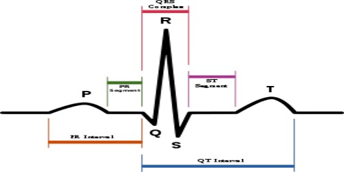
Fig: electrocardiogram
Description of the various waves of an ECG
Type of waves –
(1) Depolarization, and (2) Repolarization.
There are five consecutive waves- (1) P wave, (2) Q wave, (3) R wave, (4) S wave, (5) T wave.
P Wave
Definition: the short upward deflection is called P wave. The initial portion of the P wave is largely a reflection of right atrial depolarization and the terminal portion is largely a reflection of left atrial depolarization.
Causes of production: Atrial depolarization, therefore the passage of action current over the atria.
Duration: About 0.1 second.
Importance: Normal P indicates-
- The impulse is originating at the S.A node. So, it indicates that S.A. node is working well.
- It spreads over the atria in the usual direction.
- There is no detecting in conduction.
- The strength of contraction, the mass of atrial musculature and its nutrition are normal.
Inverted P wave: It indicates that S.A node fails to initiate impulse and the atrial muscle depolarizes by the impulse originating in A.V. node.
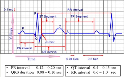
Fig: ECG – graphical representation
QRS complex
(a) Q wave: Downward deflection. The dead muscle neither conducts nor produces current, so the ECG picks up the current flowing away from this muscle, producing a strong negative deflection.
(b) R wave: Upward deflection. The R wave may be enlarged with ventricular hypertrophy, a thin chest wall or with an athletic physique.
(c) S wave: Downward deflection after R wave. It represents the late ventricular depolarization.
Cause of production: Ventricular depolarization, therefore, the passage of action current over the ventricle.
Duration: Usually less than 0.08 sec.
Importance
- Q wave indicates that impulse is generated from the septum.
- Prominent Q wave indicates old infarction.
- Absence at Q wave in infants shows congenital pliancy of a septum.
- R wave due to Rt ventricle and S wave due to Lt ventricle.
- Prolonged duration (> 0.1 sec) of R and S waves indicates the bundle of his block.
T wave – Upward broad and rounded deflection. It is normally upright, somewhat rounded, and slightly asymmetric. Its morphology will alter with breath holding and digitalis toxicity.
Cause of production:
- Ventricular repolarization.
- Contraction of the basal part of the ventricle.
Duration: 0.27 sec.
Importance: Abnormal T wave indicates serious myocardial damage.