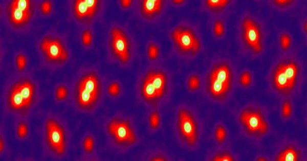
A team of Cornell University engineers created a new microscopy technique powerful enough to detect an individual atom in three dimensions — and create an image so clear that the only blurriness comes from the atom’s movement.
The technique, which, according to the study published in the journal Science, relies on an electron microscope coupled with sophisticated 3D reconstruction algorithms, does more than just break atom resolution records. According to the researchers, this is as good as microscopy gets.
“This is more than just a new record,” said lead author and Cornell engineer David Muller in a press release. “It has effectively reached a regime that will serve as an ultimate limit for resolution. We can now figure out where the atoms are in a very simple way. This opens up a slew of new measurement opportunities for things we’ve wanted to do for a long time.”
A team of Cornell University engineers developed a new microscopy technique that’s powerful enough to spot an individual atom in three dimensions — and create an image so clear that the only blurriness comes from the movement of that atom itself.
Wobbling Away
Previous attempts to image and study individual atoms resulted in blurry images because, well, getting that precise is difficult. However, scientists can now see atoms jiggle and vibrate — motion blur in the new images, they claim, is a testament to their precision rather than a technical flaw.
“With these new algorithms, we’re now able to correct for all of our microscope’s blurring to the point where the only blurring factor we have left is the fact that the atoms themselves are wobbling,” Muller said in the release, “because that’s what happens to atoms at finite temperature.”
The thrill isn’t limited to high-resolution images. The new capabilities also allow researchers to investigate previously invisible properties of materials, such as electric and magnetic fields, as well as difficult-to-detect vibrations inside crystals, in unprecedented depths. In addition, some researchers are converting the vacuum-filled interiors of electron microscopes into miniature laboratories in order to study how samples behave when exposed to liquids, gases, or varying temperatures.

Muller’s images are the latest in a series of technological advances that are ushering in a new era in what researchers can investigate using transmission electron microscopes (TEMs) — devices as tall as a room that send electron beams through samples to investigate structures as small as an atom. The machines promise to provide scientists with access to previously inaccessible details, such as the structure of fragile next-generation electronics materials and the insides of porous substances that can separate gases.
Paradigm Shift
The previous record for single-atom resolution was set in 2018 by the same team, but they could only image samples of extremely thin materials only a few atoms thick at the time. Their new technique, on the other hand, enables them to work on thicker, more realistic samples and could even be used to improve medical imaging.
Muller stated in the release, “We want to apply this to everything we do.” “Up until now, we’ve all been wearing terrible glasses. And now we have a really good couple. Why wouldn’t you take off your old glasses, put on your new ones, and wear them all the time?”
The thrill isn’t limited to high-resolution images. The new capabilities also allow researchers to investigate previously invisible properties of materials, such as electric and magnetic fields, as well as difficult-to-detect vibrations inside crystals, in unprecedented depths. In addition, some researchers are converting the vacuum-filled interiors of electron microscopes into miniature laboratories in order to study how samples behave when exposed to liquids, gases, or varying temperatures.


