Scientists have shed light on an important component of the eye: a protein found in the rod cells of the retina that allows us to see in low-light conditions. The protein relays the optical signal from the eye to the brain by acting as an ion channel in the cell membrane. If a genetic disorder disrupts a person’s molecular function, they will go blind. Scientists have deciphered the three-dimensional structure of the protein, paving the way for novel medical treatments.
PSI researchers shed light on an important component of the eye: a protein found in the rod cells of the retina that allows us to see in low light. The protein relays the optical signal from the eye to the brain by acting as an ion channel in the cell membrane. If a genetic disorder disrupts a person’s molecular function, they will go blind. Scientists have deciphered the three-dimensional structure of the protein, paving the way for novel medical treatments. The research was published in the journal Nature Structural & Molecular Biology.
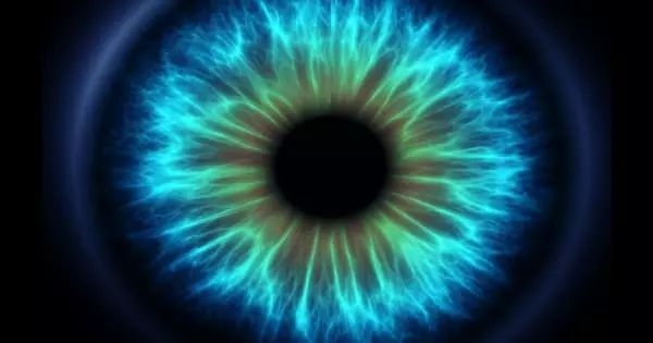
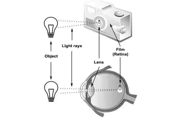
“We can see stars in the night sky because of the rod cells in our eyes,” says Jacopo Marino, a biologist at PSI’s Laboratory of Biomolecular Research. “These photocells are so sensitive to light that they can detect even a single photon reaching us from the farthest reaches of the universe – a truly incredible feat.” Our brain’s ability to eventually translate these light beams into a visual impression is due in part to cyclic nucleotide-gated (CNG) ion channels, the three-dimensional structure of which has now been illuminated by a PSI research group led by Jacopo Marino.
The scientists then used cryo-electron microscopy to reveal the ion channel’s three-dimensional structure. Unlike previous research on the structure of the ion channel, we looked at the native protein as it exists in the eye. As a result, we are much closer to understanding the true conditions that exist in living creatures.
Diane Barret
The ion channel acts as a gatekeeper, determining whether specific particles are allowed into the receptor cell’s interior. It is embedded in the rod cells’ protein-rich shell, known as the cell membrane. In the dark, the ion channel, and thus the cell’s gate, is completely open. However, when light enters the eye, it initiates a series of events in the rod cells. This eventually causes the gate to close, preventing positively charged particles such as calcium ions from entering the cell.
This electrochemical signal travels through nerve cells into the brain’s visual cortex, where it creates a visual impression, such as a flash of light. “The idea of resolving the structure of this channel dates back nearly 20 years when Gebhard Schertler and Benjamin Kaupp collaborated on this topic,” Jacopo Marino says. The new study’s authors are both co-authors.
Endurance paid off
Diane Barret, a Ph.D. student, had to first extract the channel protein from cow eyes supplied by an abattoir, which was a difficult and time-consuming process. “This was a difficult task because the protein is extremely sensitive and decomposes quickly. Furthermore, it is only found in trace amounts in the source material “Barret elaborates. It took two years to get enough protein to work with. “We were both too stubborn to just give up,” laughs Jacopo Marino. “However, in the end, that tenacity paid off.”
The scientists then used cryo-electron microscopy to reveal the ion channel’s three-dimensional structure. “Unlike previous research on the structure of the ion channel, we looked at the native protein as it exists in the eye. As a result, we are much closer to understanding the true conditions that exist in living creatures “Diane Barret expresses herself.
One of the reasons why a better understanding of the channel protein’s natural structure is important is that it will help in the development of treatments for genetic disorders with no known cure, such as retinitis pigmentosa. Photoreceptors gradually die off in this disease, leaving people blind. One possible explanation is that the body is unable to produce the CNG channel protein correctly due to a genetic defect. As a result, when light strikes the eye, the ion channel does not completely close, disrupting the cell’s electrochemical balance and causing the cells to die.
“If we could find molecules that affect the protein in such a way that the channel completely closes, we could prevent the cells from dying – and thus prevent people from going blind,” Jacopo Marino explains. Now that researchers have determined the precise structure of the protein, they can search for such molecules specifically.
Additional barrier
The protein is made up of four different subunits: three lots of subunit A and one lot of subunit B. Only in this combination is a properly functioning ion channel possible. PSI scientists demonstrate why the B subunit appears to play such an important role in their study: a side arm of the protein a single amino acid protrudes from the rest of the protein, acting as a barrier across a gateway. This narrows the channel passage to the point where no ions can pass through.
“No one expected that; it was completely unexpected,” Diane Barret says. Other narrow places, such as main gateways, which were previously thought to be the only ones, already exist in the A subunit. It is worth noting that the additional barrier is found not only in the protein from the cow’s eye, but appears to apply to all types of animals, as demonstrated by the scientists. All living creatures with an ion channel in their eye, whether crocodiles, eagles, or humans, have the same protruding amino acid at this position in the protein. Because it has been preserved so consistently throughout evolution, it must be necessary for the channel to function.