Ovule: The round shaped microscopic organs present in the placenta of the ovary which is turned to seeds after being fertilized are called ovule. An ovule is a female megasporangium where the structure of megaspores takes place. It is the structure that gives rise to and contains the female reproductive part.
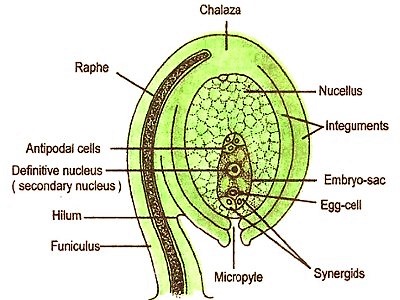
Fig: Structure of an ovule
Structure of ovule: In the flowering plant, the ovule is situated surrounded by the segment of the flower called gyanecium. Each ovule consists of nucellus surrounded by two integuments and a stalk or funiculus. A typical ovule is composed of the following organs:
- Funiculus
The connecting slender stalk between ovule and placenta is known as Funiculus. It is a stalk-like formation which represents the point of the accessory of the ovule to the placenta of the ovary. It is a stalk-like structure by which ovule is attached to the placenta.
- Hilum
The attaching point between an ovule and a funiculus is called hilum. It is the point where the body of the ovule is attached to the funiculus. It is the point of attachment of the body of the ovule with the funiculus.
- Nucellus
The main body tissue of an ovule is called nucellus. It is a mass of the parenchymatous tissue surrounded by the integuments from the outside. The nucellus provides nutrition to the developing embryo. The embryo sac is situated inside the nucellus. It is a mass of diploid cell called megasporangium. It provides sustenance in the development of embryo sac. The nucellus is a mass of cells surrounded by the integuments from the outside. Cells of nucellus have abundant reserves of food material. It is the main component of the ovule.
- Integument
One layered two outer covering- surrounding the nucellus is called Integument. They are the outer layers surrounding the ovule that provide protection to the developing embryo. Integuments form seed coats i.e. testa and tegmen. Integuments are one or two layers surrounding the ovule that endow with protection to the developing embryo.
- Micropyle
The exposed area of the anterior part of an ovule is called Micropyle. It is a narrow pore produced by the projection of integuments. It marks the point where the pollen tube enters the ovule at the time of fertilization. It is a small opening which is left by the integument in the ovule for the passage of pollen tube into the ovule. The micropyle is a pore-like structure on one side of the ovule, where integuments are absent.
- Chalaza
The area of the ovule where nucellus and integument are fused is called Chalaza. It is the based swollen part of the nucellus from where the integuments originate. The basal region of ovule from where the integuments arise is called chalaza.
- Raphe
It is the longitudinal ridge formed by the lengthwise fusion of funiculus with the body of the ovule in a typical anatropous ovule.
- Embryo sac
It is the female gametophyte which contains the egg apparatus, antipodal, and polar nuclei. Female gametophyte covered by a thin membrane is called embryo sac. It is situated in the nucellus. A mature embryo sac consists of a group of three cells at each of its ends, lying on either side of a big centrally placed nucleus.