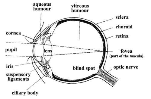
The structure and functions of human eyes are similar to a camera. We know that photograph is taken by keeping the object before a camera. Similarly, if any object is placed before the eyes, there is a picture of that image inside the eyes. The structure and functions of eyes are discussed below.
The structure of eyes

- Eye-ball: The circular object situated in the eye cavity/orbit is called the eye-ball. This ball is flattened in its front and back. It can rotate around a certain area in the eye cavity.
- Sclerotic: It is composed of white, strong and dense fibrous tissues [fig]. It determines the size of the eyes and protects the eyes and save them from any external hazards
- Cornea: It is the frontal part of sclerotic. This part of sclerotic is transparent and slightly convex at the outer side.
- Choroid: There is a deep black layer on the inner side of the sclera. This is called choroids. Due to this black layer light is not reflected internally within the eye.
- Iris: Just behind the cornea, there is an opaque diaphragm. It is called Iris. The color of the Iris may vary from person to person. Usually, the color of Iris is black, light azure or deep brown. Iris regulates the amount of light falling on the eye lens.
- Pupil: The hole at the center of the Iris is called Pupil. Through this pupil, light enters the eyes.
- Eye lens: It is the most important part of the eye. It is situated just behind the pupil. It is made of transparent organic substances. Its radius of curvature of the back side is greater than that of the front side. This lens is hooked in the eyeball by ciliary muscles and suspensory ligaments. Due to the contractions and suspension of these muscles and ligaments, the curvature of the lens changes. It ultimately changes the focal length. To see different objects of near and distance, we need to adjust our focal length.
- Retina: Just behind the eye lens, there is a layer of semi-transparent light-sensitive membrane on the innermost side of the eyeball. It is called the retina. It is made of some nerve-fibers called rods and cones. These nerve-fibers are connected to the eye-nerves. When light falls on the retina, it creates a kind of excitement in nerve-fibers. Hence. a sensation of vision is created in the brain.


