Nanoscale devices are 100 to 10,000 times smaller than human cells. They have the same size as large biological molecules (“biomolecules”) like enzymes and receptors. Hemoglobin, the molecule that transports oxygen in red blood cells, has a diameter of about 5 nanometers.
Nanoscale devices less than 50 nanometers in size can easily enter most cells, while those less than 20 nanometers in size can move out of blood vessels as they circulate throughout the body. Nanoscale devices, due to their small size, can easily interact with biomolecules on both the surface and inside cells. They have the potential to detect disease and deliver treatment in previously unimagined ways by gaining access to so many areas of the body.
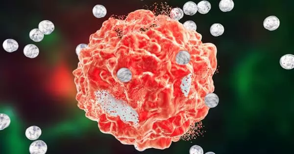
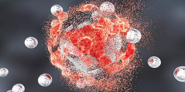
A multi-institutional research team created nanoparticles that can communicate with cancer cells and slow their growth. The research described in a new paper published in Advanced Materials has revealed a novel framework for the potential development of drug-free cancer therapies.
There are currently no nanoparticle formulations that target diseased cells in the body optimally. A small percentage of nanoparticles reach these cells, with the majority accumulating in mononuclear phagocytic cells.
Cancer cells take up materials from their environment, and they also secrete factors that help them degrade surrounding tissue in order to spread and metastasize. We created particles that respond to both of these characteristics by aggregating into clusters that cancer cells actively take up.
Richard Huang
The research team, led by scientists at the Advanced Science Research Center at the Graduate Center, CUNY (CUNY ASRC), was able to design nanoparticles that are activated to self-assemble when they come into contact with cancer cells and send messages instructing the cells to slow their growth. Because the nanoparticles only communicate with cancer cells, the healthy cells around them are unaffected.
“Cancer cells take up materials from their environment, and they also secrete factors that help them degrade surrounding tissue in order to spread and metastasize,” said Richard Huang, the paper’s lead author and a Ph.D. student at the Graduate Center, CUNY (GC CUNY), as well as a researcher with the CUNY ASRC Nanoscience Initiative and the Center for Discovery and Innovation at CUNY’s City College of New York (CCNY). “We created particles that respond to both of these characteristics by aggregating into clusters that cancer cells actively take up. Once inside, they appear to be able to reduce the metabolic activity of cancer cells, thereby slowing their growth.”
The cells secrete an abnormally large amount of the matrix metalloproteinase-9 (MMP-9) enzyme, which breaks down the collagen that holds healthy tissue together, which is one reason cancer progression is difficult to control. This feature was exploited by the research team against cancer cells. This is accomplished by creating nanoparticles that, when activated by MMP-9, begin assembling into large aggregates in the vicinity of the cells. These aggregates are engulfed by the cells, and their size causes physical distress to the cancer cells, reducing their ability to proliferate and survive.
The researchers were able to use confocal reflection microscopy to visualize the nanoparticle aggregates inside the cancer cells in real-time, which is one of the study’s highlights. “This label-free, live imaging technique allowed us to get a better look at when and where the aggregates formed, as well as how cancer cells respond to the particles at the sub-cellular level,” said Ye He, director of the CUNY ASRC Neuroscience Initiative’s Live Imaging and Bioenergetics Facility.
At the nanoscale, biological processes, both necessary for life and those that lead to cancer, take place. As a result, we are made up of a slew of biological nano-machines. Nanotechnology allows researchers to study and manipulate macromolecules in real time, at the earliest stages of cancer progression. Nanotechnology has the potential to provide rapid and sensitive detection of cancer-related molecules, allowing scientists to detect molecular changes that occur in only a small percentage of cells. Nanotechnology has the potential to create completely novel and highly effective therapeutic agents.
“We wanted to see if we could use relatively simple peptide design to create nanoparticles that could produce robust self-assembly in biological media and have an effect on cancer cells,” said Rein Ulijn, director of the CUNY ASRC Nanoscience Initiative and the study’s principal investigator. “While this is early-stage research, the findings offer exciting prospects for a drug-free therapeutic treatment that could be extremely useful for cancer patients who have developed drug resistance as well as for extending the lives of people with metastatic cancer.”
More research is needed to fully understand the therapeutic potential of the team’s discovery.