Scientists have successfully implanted a bioinspired steerable catheter into an animal’s brain. Early-stage research examined the delivery and safety of the new implantable catheter design in two sheep to determine its utility in diagnosing and treating brain diseases.
If the platform is proven to be effective and safe for human use, it could simplify and reduce the risks associated with diagnosing and treating disease in the deep, delicate recesses of the brain. It could help surgeons see deeper into the brain to diagnose disease, deliver more precise treatment like drugs and laser ablation to tumors, and better deploy electrodes for deep brain stimulation in conditions like Parkinson’s and epilepsy.
“The brain is a fragile, complex web of tightly packed nerve cells that each have their part to play,” said senior author Professor Ferdinando Rodriguez y Baena of Imperial’s Department of Mechanical Engineering. “When disease arises, we want to be able to navigate this delicate environment to precisely target those areas without harming healthy cells.”
“Our new precise, minimally invasive platform improves on currently available technology and, if proven safe and effective, could enhance our ability to safely and effectively diagnose and treat diseases in people.”
Developed as part of the Enhanced Delivery Ecosystem for Neurosurgery in 2020 (EDEN2020) project, the findings are published in PLOS ONE.
Our findings may have far-reaching implications for minimally invasive, robotic-assisted brain surgery. We hope it will contribute to the improvement of the safety and effectiveness of current neurosurgical procedures requiring precise deployment of treatment and diagnostic systems, such as in the context of localised gene therapy.
Professor Ferdinando Rodriguez
Stealth surgery
The platform improves on existing minimally invasive, or “keyhole,” surgery, in which surgeons insert tiny cameras and catheters into the body through small incisions. It consists of a soft, flexible catheter to avoid damaging brain tissue during treatment and an artificial intelligence (AI)-enabled robotic arm to assist surgeons in navigating the catheter through brain tissue.
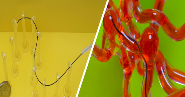
The catheter, which was inspired by the organs used by parasitic wasps to lay eggs in tree bark, is made up of four interlocking segments that slide over one another to allow for flexible navigation.
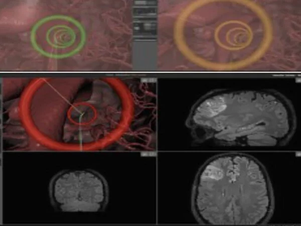
It connects to a robotic platform that combines human input and machine learning to carefully steer the catheter to the disease site. Surgeons then deliver optical fibres via the catheter so they can see and navigate the tip along brain tissue via joystick control.
To guide the catheter with pinpoint accuracy, the AI platform learns from the surgeon’s input and contact forces within brain tissues. In comparison to traditional ‘open’ surgical techniques, the new approach may eventually help to reduce tissue damage during surgery, as well as improve patient recovery times and length of post-operative hospital stays.
Surgeons use deeply penetrating catheters to diagnose and treat disease while performing minimally invasive brain surgery. However, current catheters are rigid and difficult to precisely place without the use of robotic navigational tools. The inflexibility of the catheters, combined with the intricate, delicate structure of the brain, means catheters can be difficult to precisely place, posing risks to this type of surgery.
To test their platform, the researchers deployed the catheter in the brains of two live sheep at the University of Milan’s Veterinary Medicine Campus. The sheep were given pain relief and monitored for 24 hours a day over a week for signs of pain or distress before being euthanized so that researchers could examine the structural impact of the catheter on brain tissue.
They found no signs of suffering, tissue damage, or infection following catheter implantation.
Lead author Dr. Riccardo Secoli, also from Imperial’s Department of Mechanical Engineering, said: “Our analysis showed that we implanted these new catheters safely, without damage, infection, or suffering. If we achieve equally promising results in humans, we hope we may be able to see this platform in the clinic within four years.
“Our findings may have far-reaching implications for minimally invasive, robotic-assisted brain surgery. We hope it will contribute to the improvement of the safety and effectiveness of current neurosurgical procedures requiring the precise deployment of treatment and diagnostic systems, such as in the context of localized gene therapy.”
Professor Lorenzo Bello, co-author of the study from the University of Milan, stated: “One of the most significant limitations of current MIS is that if you want to reach a deep-seated site through a burr hole in the skull, you are limited to a straight-line trajectory. The rigid catheter’s accuracy within the shifting tissues of the brain, as well as the tissue deformation it can cause, are its limitations. We’ve discovered that our steerable catheter can overcome the majority of these limitations.”