Tissue imaging is an important aspect of translational research that bridges the gap between basic laboratory science and clinical science to improve cancer understanding and aid in the development of new therapies. To fully analyze images, scientists should ideally use an application that allows multiple images to be viewed at the same time.
Moffitt Cancer Center researchers describe a new open-source software program they developed that allows users to view many multiplexed images at the same time in an article published in the journal Patterns.
Over the last decade, there have been significant advancements in cancer research methods, including new techniques for studying tissue samples. Machines, for example, can now be programmed to stain hundreds of slides at the same time, or up to 1,000 different tissue sample cores can be placed on a single slide and stained for biomarkers at the same time.
We will enhance Mistic to use biologically meaningful regions of interest from the multiplexed image to render the overall image t-SNE. We also have plans to augment Mistic with other visualization software and build a cross-platform viewer plugin to improve the adoption, usability and functionality of Mistic in the biomedical research community.
Sandy Anderson
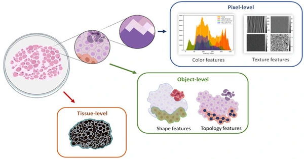
With the introduction of these approaches comes a plethora of opportunities to generate new data and information. To view and study the cancer biomarkers, tissue architecture, and cellular interactions among these samples, computational modeling and software are required due to the magnitude of this information and the complexity of cancer itself.
While working on a project, researchers in Moffitt’s Integrated Mathematical Oncology Department (IMO) realized that the currently available software for image viewing was insufficient for their needs.
“We were curious about the underlying spatial patterns between tumor and immune cells, as well as how tumors were organized. This required us to compare multiple images at the same time, and we discovered that there was no free or commercial software that could do so “Moffitt’s lead author and applied research scientist, Sandhya Prabhakaran, Ph.D.
The IMO team decided to develop a software program that would allow them to view multiple images at the same time and extract data through additional analyses that could be used for a variety of purposes, such as identifying biomarkers and understanding tissue architecture and the spatial organization of different cell types.
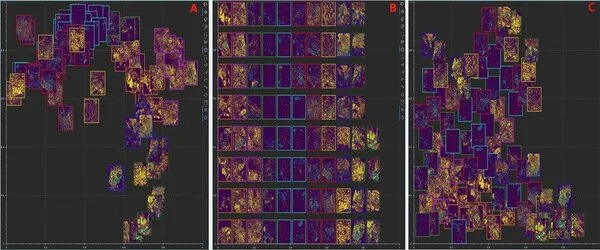
Mistic, their program, takes information from multidimensional images and uses dimensionality reduction methods called t-distributed stochastic neighbor embedding (t-SNE) to abstract each image to a point in reduced space. Mistic is an open-source software that works with images from Vectra, CyCIF, t-CyCIF, and CODEX.
The researchers describe the development of Mistic as well as some of the applications that it could be used for in their publication. They demonstrated, for example, that the software could be used to analyze 92 images from patients with non-small cell lung cancer and determine how biomarkers cluster across patients with different treatment responses. Another study used Mistic in conjunction with statistical analysis to assess the spatial colocalization and coexpression of immune cell markers in 210 endometrial cancer samples.
The team is enthusiastic about Mistic’s potential applications and has plans to improve the software.
“We will enhance Mistic to use biologically meaningful regions of interest from the multiplexed image to render the overall image t-SNE. We also have plans to augment Mistic with other visualization software and build a cross-platform viewer plugin to improve the adoption, usability and functionality of Mistic in the biomedical research community,” said Sandy Anderson, Ph.D., author and chair of Moffitt’s IMO Department.
In addition to Mistic, the Patterns featured the IMO team in a People of Data article titled “Developing tools for analyzing and viewing multiplexed images.” Here, the IMO team gets to introduce themselves, discuss their research passion and the challenges and opportunities relevant to imaging in mathematical oncology.