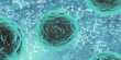
A pollen particle revealing the nanofoam within, or a diatom revealing the distinct geometric features within A team led by CFEL scientists Saa Bajt and Henry Chapman was able to scan these structures without destroying them using high-energy X-rays from DESY’s PETRA III synchrotron light source.
Their new technique produces high-resolution X-ray photographs of dried biological material that has not been frozen, coated, or otherwise altered—all with little to no sample damage. This technology, which is also used for airport baggage scanning, may provide images with nanometre resolution of the substance.
The particular technology permits imaging to be conducted at less than 1% of the specimen’s X-ray damage threshold by using high-energy X-rays that are sharply focussed using a set of innovative diffractive lenses. The findings, which show that this technology is a promising tool for brighter next-generation light sources like the planned upgrade project PETRA IV, were published in the journal Light: Science & Applications.
X-ray radiation interacts with biological material in a variety of ways, the majority of which are determined by the light’s energy and intensity. At the same time, radiation damage, which can range from minor structural changes to complete sample disintegration, is a limiting issue in X-ray imaging of biological material.
At low energies, the X-rays are predominantly absorbed by the atoms in the sample, causing their electrons to absorb the energy and spring out of the atoms, causing damage to the sample. Images created with these low-energy X-rays map out the sample’s radiation absorption. At higher energies, absorption becomes less likely, and a phenomenon known as elastic scattering takes place, in which X-ray photons “bounce” off of materials like billiard balls without depositing their energy.
This interaction is used in techniques such as crystallography and ptychography. Nonetheless, absorption can occur, implying that sample deterioration occurs regardless. However, there is a third interaction: Compton scattering, in which the X-rays leave only a trace of their energy in the target material. Compton scattering had been widely overlooked as a feasible method of X-ray microscopy since it necessitates even higher X-ray energy, where no suitable high-resolution lenses existed until recently.
“We used Compton scattering and figured out that the amount of energy deposited into a sample per a number of photons that you can detect is lower than using these other methods,” says Chapman, who is a leading scientist at DESY, a professor at Universität Hamburg, and the inventor of various X-ray techniques at synchrotrons and free-electron lasers.
The minimal dose in the sample provided a hurdle in creating acceptable lenses. High-energy X-rays penetrate through all materials and are only slightly refracted or bent when focused. Bajt, a group leader at CFEL, spearheaded attempts to create a novel type of refractive lens known as multilayer Laue lenses. Over 7300 nanometer-thin alternating layers of silicon carbide and tungsten carbide were employed to produce a holographic optical element thick enough to efficiently concentrate the X-ray beam in these novel optics.
The scientists photographed a range of biological materials using this lens setup and the PETRA III beamline P07 at DESY by monitoring Compton scattering data as the sample went through the focussed beam. This scanning microscopy method necessitates the employment of an extremely bright source—the brighter, the better—that is focused on a spot that specifies the image resolution.
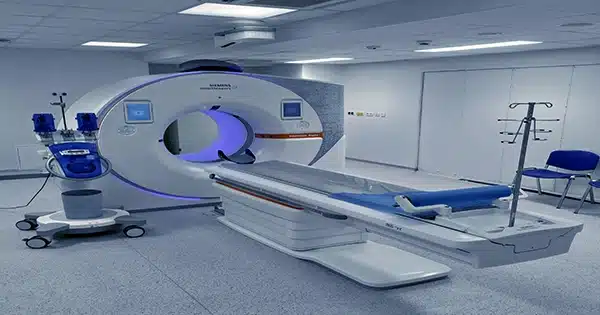
PETRA III is one of the few synchrotron radiation facilities in the world that is bright enough at high X-ray energy to take images in a reasonable amount of time. With the projected PETRA IV facility, the technology could attain its full potential.
The team utilized a cyanobacterium, a diatom, and even a pollen grain picked just outside the lab as samples (“a very local specimen,” Bajt laughs) and obtained a resolution of 70 nanometers for each.
Furthermore, when compared to images acquired from the same pollen sample using a conventional coherent-scattering imaging approach at 17 keV, Compton X-ray microscopy achieved a similar resolution with a 2000-fold lower X-ray dose. “When we re-examined the specimens using a light microscope after the experiment, we could not see any trace of where the beam had come into contact with them,” she says, implying that no radioactive damage was left behind.
“These results could be even better,” says Chapman. “Ideally, an experiment like this would use a spherical detector because the X-rays emitted by the sample travel in all directions.” In that sense, it’s similar to a particle physics collision experiment in that data must be collected from all directions.”
Furthermore, Chapman pointed out that the cyanobacteria image is very featureless in comparison to the others. However, the statistics show that with higher brightness, such as that of the proposed PETRA IV upgrade, individual organelles, and even three-dimensional structures will be visible—up to a resolution of 10 nm without causing harm. “Really, the only limitation of this technique was not the nature of the technique itself, but rather the source, namely its brightness,” Bajt explains.
With a brighter source, the technology might be used to image whole unsanctioned cells or tissue, complementing cryo-electron microscopy and super-resolution optical microscopy, or to follow nanoparticles within a cell, such as to directly observe drug delivery. Compton scattering’s properties make it perfect for non-biological applications such as studying the physics of battery charging and discharging.
“There hasn’t been anything like this technique in the literature yet,” Bajt says, “so there is much to explore going forward.”