A New Progress to Ultrasound Imaging
Conventional ultrasonic imaging requires acoustic scanning over a target object using a piezoelectric transducer array, followed by signal processing to reconstruct the image. The researchers have developed a receiver that incorporates a piezoelectric crystal and an organic light-emitting diode (OLED). “Conventional ultrasound devices have a receiver that detects ultrasonic waves and converts them into an electrical signal, which is then sent to a computer that processes the signal and converts it into an image,” says Xiaoning Jiang, co-corresponding author of a paper on the work and a Duncan Distinguished Professor of Mechanical and Aerospace Engineering at NC State. “Conventional ultrasound imaging probes can cost upward of $100,000 because they contain thousands of transducer array elements, which drives up manufacturing costs.” “We’ve created a device that effectively eliminates the electrical signal processing altogether.” When an ultrasonic wave hits the crystal, it produces a voltage, which causes the OLED to light up. In other words, the image appears on the OLED screen, which is built into the receiver itself.
The researchers are interested in collaborating with industry partners to explore commercial applications. Specifically, the researchers have developed a receiver that incorporates a piezoelectric crystal and an organic light-emitting diode (OLED). Ultrasound imaging (sonography) uses high-frequency sound waves to view inside the body. When an ultrasonic wave hits the crystal, it produces a voltage, which causes the OLED to light up. In other words, the image appears on the OLED screen, which is built into the receiver itself. Because ultrasound images are captured in real-time, they can also show the movement of the body’s internal organs as well as blood flowing through the blood vessels.
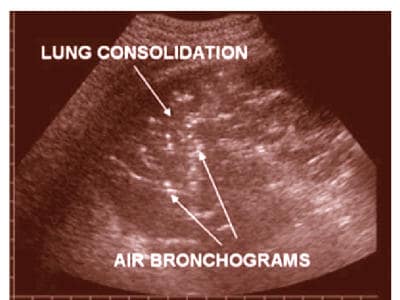
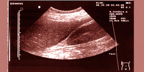
Fig: Ultrasound imaging of the patient’s lung.
Ultrasound imaging uses sound waves to produce pictures of the inside of the body. “Our prototype is a proof-of-concept, so we designed it with an OLED array that is 10 pixels by 10 pixels; the resolution isn’t great,” says Franky So, co-corresponding author of the study. Ultrasound has several advantages which make it ideal in numerous situations, in particular, studies of the function of moving structures in real-time. “However, I can easily make it 500 pixels by 500 pixels, boosting the resolution substantially.” So is the Walter and Ida Freeman Distinguished Professor of Materials Science and Engineering at NC State.
Conventional ultrasound displays the images in thin, flat sections of the body. “Conventional ultrasound imaging probes can cost upward of $100,000 because they contain thousands of transducer array elements, which drives up manufacturing costs,” So says. “We can make ultrasound receiver-display units for $100 or so.” Advancements in ultrasound technology include three-dimensional (3-D) ultrasound that formats the sound wave data into 3-D images.
“This is really a completely new field for ultrasound, so we’re only beginning to explore the potential applications,” Jiang says. It can be used to examine many parts of the body, such as the abdomen, heart and blood vessels, breasts, muscles, carotid arteries, and female reproductive system including pregnancy and prenatal diagnostics. “However, there are obvious near-term applications, such as non-destructive testing, evaluation, and inspections in the context of structural health monitoring.” Ultrasound is based on sonar and uses a machine with a computer processor to create Ultrasound images. Returning sound waves, or echoes, are processed through the computer and converted into images.