
Fetal MRI imaging can help clinicians provide better care for children both before and after birth by more precisely revealing any head, neck, thoracic, abdominal, and spinal malformations in unborn babies. A large multidisciplinary study led by King’s College London with Evelina London Children’s Hospital, Great Ormond Street Hospital, and UCL discovered that MRI scanning can more precisely define and detect head, neck, thoracic, abdominal, and spinal malformations in unborn babies.
The team of researchers and clinicians demonstrate how MRI scanning can show malformations in great detail, including their effect on surrounding structures, in the study, which was published today in the Lancet Child and Adolescent Health. They emphasize that MRI is a completely safe procedure for pregnant women and their babies.
They claim that the work is invaluable to both clinicians caring for babies before birth and teams planning care for the baby after birth.
MRI imaging is more adept at detecting fetal abnormalities than conventional ultrasound imaging. The work is invaluable both to clinicians caring for babies before they are born and for teams planning the care of the baby after delivery.
Recent studies have focused on correcting for fetal movement in fetal brain MRI and, more recently, imaging the fetal heart. However, there is an increasing demand for MRI to evaluate the entire fetus, and researchers from King’s College London’s School of Biomedical Engineering & Imaging Sciences at Evelina London Children’s Hospital have recently developed a post-acquisition pipeline to motion correct and volume reconstructs images of the entire fetal body.
Ultrasound is still considered the gold standard for fetal imaging, but it can fall short at times and for certain needs. MRI is also safe for pregnant women and their unborn children, according to the team led by Mary Rutherford, professor in the School of Biomedical Engineering & Imaging Sciences. According to the lead researcher, Professor Mary Rutherford of the School of Biomedical Engineering & Imaging Sciences, ultrasound remains the gold standard for fetal screening and is indeed complementary to these optimized MRI approaches for evaluating fetal body abnormalities.
“Up until now, ultrasound was the modality of choice for diagnosing those anomalies. However, ultrasound’s ability to define the most detailed anatomy is sometimes limited. MRI scanning has the potential to more precisely define malformations, which could aid clinicians in their care planning and parent counseling.”
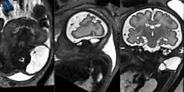
MRI is commonly used to classify fetal brain anomalies. Although its use in fetal body anomalies is less common, advances have validated its role in the antenatal investigation of several conditions, including the investigation of fetuses with spina bifida: imaging of the fetal brain as well as the spinal cord is an important factor in determining which patients may benefit from fetal surgery.
For fetal neck masses, MRI clearly outperforms conventional ultrasound in terms of assessing tumor extension and providing a 3D visualisation of the tumor’s relationship to the airway. MRI may also be superior to ultrasound in distinguishing between normal and abnormal lung tissue, as well as in making other diagnoses such as diaphragmatic hernia–especially in late gestation, when ultrasound is difficult to use.
New approaches to imaging the fetal body with MRI allow for both motion correction and volume reconstruction of body organs and defects. According to the researchers, this improves visualisation and, as a result, the detection and characterisation of abnormalities.
The project brought together surgeons, fetal medicine specialists, radiologists, and physicists to review the use of magnetic resonance imaging to investigate conditions in the unborn baby; this approach is already in use at Evelina London, which is part of Guy’s and St Thomas’ NHS Foundation Trust. There are also videos and images to view.
The team explained that in some cases, MRI clearly outperforms ultrasound. It can, for example, clearly assess tumor extension and provide 3D visualization for fetal neck masses as they relate to the airway, as well as provide better differentiation between normal and abnormal lung tissue. Furthermore, it can provide more information than ultrasound for diaphragmatic hernia, especially late in pregnancy. Ongoing research is aimed at developing a fully automated process suitable for clinical translation and wider dissemination into clinical practice.