
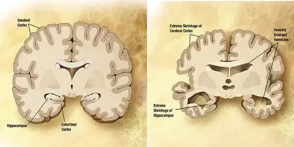
The neurons most affected in Alzheimer’s disease are typically found in specific brain regions involved in memory and cognition. The hippocampus, which is important in memory formation, is one of the first and most severely affected areas in Alzheimer’s disease. The entorhinal cortex, which is linked to the hippocampus, is also vulnerable to damage.
Researchers have identified neurons in the brain that may contribute to some of the earliest symptoms of Alzheimer’s disease, making it a promising target for potential new Alzheimer’s drugs. One of the hallmarks of Alzheimer’s disease is neurodegeneration, or the gradual loss of neuron function. It does not, however, affect all parts of the brain equally.
The mammillary body, a part of the hypothalamus, is one of the first brain regions to show neurodegeneration in Alzheimer’s disease. MIT researchers have identified a subset of neurons within this body that are most vulnerable to neurodegeneration and hyperactivity in a new study. They also discovered that this damage causes memory impairments.
According to the researchers, the findings suggest that this region may contribute to some of the earliest symptoms of Alzheimer’s disease, making it a good target for potential new drugs to treat the disease.
If we could identify specific molecular properties of classes of neurons that are predisposed to dysfunction and degeneration, then we would have a better understanding of neurodegeneration. This is clinically important because we could find ways to therapeutically target these vulnerable populations and potentially delay the onset of cognitive decline.
Mitchell Murdock
“It is fascinating that only the lateral mammillary body neurons, not those in the medial mammillary body, become hyperactive and undergo neurodegeneration in Alzheimer’s disease,” says Li-Huei Tsai, director of MIT’s Picower Institute for Learning and Memory and the senior author of the study.
In a study of mice, the researchers showed that they could reverse memory impairments caused by hyperactivity and neurodegeneration in mammillary body neurons by treating them with a drug that is now used to treat epilepsy.
Former MIT postdoc Wen-Chin (Brian) Huang and MIT graduate students Zhuyu (Verna) Peng and Mitchell Murdock are the lead authors of the paper, which appears today in Science Translational Medicine.
Predisposed to degeneration
Neurodegeneration occurs as Alzheimer’s disease progresses, along with the accumulation of amyloid beta plaques and misfolded Tau proteins, which form tangles in the brain. One unanswered question is whether this neurodegeneration affects all neurons equally or whether certain types of neurons are more vulnerable.
“If we could identify specific molecular properties of classes of neurons that are predisposed to dysfunction and degeneration, then we would have a better understanding of neurodegeneration,” Murdock says. “This is clinically important because we could find ways to therapeutically target these vulnerable populations and potentially delay the onset of cognitive decline.”
In a 2019 study using a mouse model of Alzheimer’s disease, Tsai, Huang, and others found that the mammillary bodies – a pair of structures found on the left and right underside of the hypothalamus — had the highest density of amyloid beta. These bodies are known to be involved in memory, but their exact role in normal memory and in Alzheimer’s disease is unknown.
The researchers used single-cell RNA-sequencing to learn more about the function of the mammillary body, which can reveal the genes that are active within different types of cells in a tissue sample. The researchers used this method to identify two major populations of neurons: one in the medial mammillary body and one in the lateral mammillary body. Genes related to synaptic activity were found to be highly expressed in lateral neurons, and these neurons had higher spiking rates than medial mammillary body neurons.
Based on these differences, the researchers speculated that lateral neurons might be more vulnerable to Alzheimer’s disease. They investigated this question by studying a mouse model with five genetic mutations linked to early-onset Alzheimer’s disease in humans. The researchers discovered that these mice had significantly higher levels of hyperactivity in lateral mammillary body neurons than healthy mice. However, no such differences were found in the medial mammillary body neurons of healthy mice or the Alzheimer’s model.
The researchers discovered that this hyperactivity manifested itself very early, around two months of age (the equivalent of a young human adult), before amyloid plaques formed. As the mice aged, the lateral neurons became even more hyperactive, and they were also more susceptible to neurodegeneration than the medial neurons.
“We believe the hyperactivity is related to memory circuit dysfunction as well as a cellular progression that may lead to neuronal death,” Murdock says.
The Alzheimer’s mouse model showed impairments in the formation of new memories, but when the mice were given a drug that reduces neuronal hyperactivity, their performance on memory tasks improved significantly. This drug, known as levetiracetam, is used to treat epileptic seizures and is also being tested in Alzheimer’s patients to treat epileptiform activity (hyperexcitability in the cortex, which increases the risk of seizures).
Comparing mice and humans
The researchers also studied human brain tissue from the Religious Orders Study/Memory and Aging Project (ROSMAP), a longitudinal study that has tracked memory, motor, and other age-related issues in older people since 1994. Using single-cell RNA-sequencing of mammillary body tissue from people with and without Alzheimer’s disease, the researchers found two clusters of neurons that correspond to the lateral and medial mammillary body neurons they found in mice.
Similar to the mouse studies, the researchers discovered hyperactivity signatures in the lateral mammillary bodies of Alzheimer’s tissue samples, such as overexpression of genes encoding potassium and sodium channels. They also discovered higher levels of neurodegeneration in the lateral neuron cluster than in the medial neuron cluster in those samples.
Other studies of Alzheimer’s patients have discovered a loss of mammillary body volume early in the disease, as well as plaque deposition and altered synaptic structure. According to the researchers, all of these findings suggest that the mammillary body could be a good target for potential drugs that could slow the progression of Alzheimer’s disease.