The segmentation of three-dimensional brain region contours is critical for the analysis of various brain structures, and advanced approaches are constantly emerging in the field of neurosciences. Whole-brain images can now be acquired at the cellular level thanks to advancements in high-resolution micro-optical imaging.
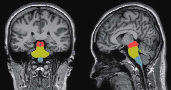
Researchers from King’s College London’s School of Biomedical Engineering & Imaging Sciences automated brain MRI image labeling, which is required to teach machine learning image recognition models, by deriving important labels from radiology reports and accurately assigning them to the corresponding MRI examinations. More than 100,000 MRI examinations can now be labeled in less than a half-hour.
This is the first study, published in European Radiology, that allows researchers to label complex MRI image datasets at scale. According to the researchers, manually labeling more than 100,000 MRI examinations would take years.
Researchers have automated brain MRI image labeling, needed to teach machine learning image recognition models, by deriving important labels from radiology reports and accurately assigning them to the corresponding MRI examinations.
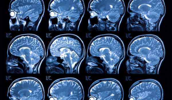
To achieve the best possible performance in image recognition tasks, deep learning typically requires tens of thousands of labeled images. This represents a barrier to the development of deep learning systems for complex image datasets, particularly MRI, which is essential for detecting neurological abnormalities.
The development of high-performing deep-learning models for image recognition tasks in radiology necessitates the collection of large-labeled datasets, a difficult and time-consuming task that has hampered algorithm development and adoption. The research team attempted to address this issue by developing a deep learning-based report classifier capable of performing automated labeling of these neuroradiology studies based on their reports.
Dr. Tom Booth, a senior author from King’s College London’s School of Biomedical Engineering & Imaging Sciences, stated: “We have greatly facilitated future deep learning image recognition tasks by overcoming this bottleneck, and this will almost certainly hasten the arrival of automated brain MRI readers into the clinic. The opportunity for patient benefit from, ultimately, timely diagnosis is enormous.”
Dr. Booth described their validation as “unusually robust.” Instead of evaluating their model performance on unseen radiology reports, they also evaluated it on unseen images. “While this may seem obvious, it has been difficult to achieve in medical imaging because it necessitates a large team of expert radiologists. Fortunately, our team is an ideal blend of clinicians and scientists “Dr. Booth stated.
Lead Author, Dr David Wood from the School of Biomedical Engineering & Imaging Sciences said: “This study builds on recent breakthroughs in natural language processing, particularly the release of large transformer-based models such as BERT and BioBERT which have been trained on huge collections of unlabeled text such as all of English Wikipedia, and all PubMed Central abstracts and full-text articles; in the spirit of open-access science, we have also made our code and models available to other researchers to ensure that as many people benefit from this work as possible.”
According to the authors, while one barrier has now been overcome, further challenges will be, first, to perform the deep learning image recognition tasks, which also have multiple technical challenges; and second, once this is accomplished, to ensure that the developed models continue to perform accurately across different hospitals using different scanners.
Dr. Booth stated: “This research was made possible by a large group of experts who are working on these issues. There is a large group of organizers and facilitators who are equally important in carrying out this research. Obtaining clean data from multiple hospitals across the UK is a critical first step toward overcoming the next challenges. We are conducting an NIHR portfolio-adopted study across the UK to collect brain MRI data prospectively for this purpose.”
They will face additional challenges in performing deep learning image recognition tasks to ensure that the developed models continue to perform accurately across different hospitals using different scanners.
In the next stage of their research, the team is tackling the challenge of performing deep-learning image recognition tasks, which also present a number of technical challenges. Once this is accomplished, the authors stated that they will ensure that the developed models continue to perform accurately across different hospitals and scanners.