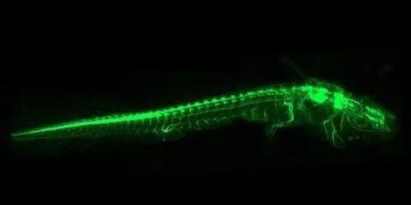
Deep learning has shown great potential in improving the speed and accuracy of whole-body imaging of small animals. With the help of deep learning algorithms, it is possible to acquire high-quality images of small animals in a much shorter time frame than traditional imaging techniques.
A research team presents technology that uses deep learning to improve photoacoustic computed tomography. After a flash of lightning, the sound of thunder takes a few moments to reach our ears. The photoacoustic (PA) effect causes materials near lightning to instantly expand as the lightning’s optical energy is absorbed and converted into thermal energy.
Photoacoustic computed tomography (PACT) has become a premier preclinical and clinical imaging modality for taking images inside the body without the use of a contrast medium by utilizing this PA effect. However, without such hardware, its low-quality images, which can be improved with multiple ultrasound sensors and a multi-channel data acquisition (DAQ) system, result in higher costs and slower imaging speed.
While deep learning has been used in previous studies to improve resolution, this is the first study in the world to apply deep learning to the three-dimensional multiparametric PACT system.
A POSTECH research team – consisting of Professor Chulhong Kim and Ph.D. candidate Seongwook Choi (Department of Convergence IT Engineering), Professor Seungchul Lee and Ph.D. candidate Soo Young Lee (Department of Mechanical Engineering), and Dr. Jinge Yang (Department of Electrical Engineering) — has presented a deep-learning approach to achieve faster and high-resolution imaging for the PACT system. The finding, the first in the world, was recently published in Advanced Science.
While deep learning has been used in previous studies to improve resolution, this is the first study in the world to apply deep learning to the three-dimensional multiparametric PACT system. The researchers demonstrated that high-resolution, high-speed, and real-time imaging of tissues in the heart, kidney, and brain, as well as whole-body imaging of animals, is possible. They’ve also demonstrated for the first time that deep learning can be used in pharmacokinetics, which involves injecting drugs into blood vessels to track how they spread throughout the body, and functional imaging, which measures the oxygen saturation of each tissue.

The researchers also confirmed that an artificial neural network trained on animals can be applied to humans through this study. It’s also significant that they simplified the hardware without sacrificing speed or quality because the artificial neural network operates independently of the optical wavelength used to train it. The research team anticipates that with the publication of the findings, the PACT technology will be widely applicable in a variety of environments by achieving high-resolution and high-speed images regardless of hardware specifications. This study was chosen as the back-cover paper in the latest issue of Advanced Science because of its significance.
One of the most common deep learning-based techniques used for whole-body imaging is called compressed sensing MRI (CS-MRI). This technique uses a deep learning algorithm to predict the missing k-space data in the MRI scan, allowing for a significant reduction in imaging time while maintaining image quality. In addition, CS-MRI has shown promising results in reducing motion artifacts, making it an ideal technique for imaging small animals that tend to move frequently.
Overall, deep learning-based techniques have the potential to revolutionize whole-body imaging of small animals, allowing for faster and sharper imaging with less motion artifacts. As deep learning technology continues to evolve, it is likely that we will see even more advanced techniques and algorithms being developed to further improve imaging capabilities.


