A team of researchers from the University of Kentucky and Cincinnati Children’s Hospital is delving further into the science of how spiny mice recover missing tissue and utilizing what they discover to stimulate regeneration in other types of mice — breakthroughs that may one day be applied to people.
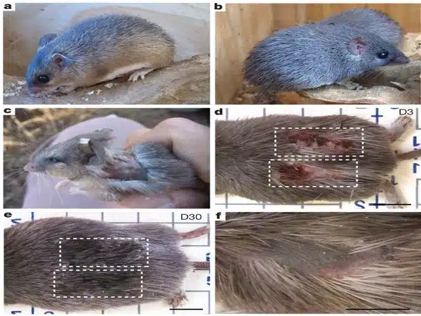
Adult laboratory mice recover injuries using scar tissue, but spiny mice have the unique capacity to regenerate missing skin and musculoskeletal structures in their bodies. Ashley W. Seifert, Ph.D., an associate professor in the Department of Biology at the UK College of Arts and Sciences, and his research team have pioneered the use of spiny mice and other animal models to understand how complex tissue regenerates, bridging the gap between regenerative biology and medicine.
In April 2023, Science Advances published a paper from Seifert’s lab on the cellular response to injury during tissue recovery. The article highlighted how ERK signaling functions as a crucial switch balancing the healing response, as well as how enhancing and sustaining this sort of signaling in laboratory mice can result in a regenerative response rather than scarring. Their most recent study, published in Developmental Cell, sheds fresh light on how certain immune cells respond to injury and direct tissue regeneration.
Our study shows that spiny mouse macrophages release distinct proteins that are partially responsible for the reformation of specialized tissues at the site of injury and for protecting cells from stress. Overall, our experiments point towards macrophages establishing a tissue microenvironment conducive for regeneration.
Ashley W. Seifert
The researchers looked at macrophages, a kind of immune cell that plays an important role in controlling the inflammatory response by defending wounded tissue against pathogens while also aiding tissue repair in both spiny and lab mice.
“In our study, we used a dual-species system to dissect macrophage phenotypes between two modes of healing: tissue regeneration or scar tissue formation,” said Seifert. “While it’s still unclear the extent to which macrophages ultimately control different healing trajectories, we observed subtle and distinct macrophage signatures that aligned specifically with regeneration.”
Using an identical injury in both kinds of rodents, the researchers studied signals released by macrophages that communicate to nearby cells, telling them to rebuild all the tissues that are missing. They also noted the characteristics, or phenotypes, of the cells during those processes.
“Our study shows that spiny mouse macrophages release distinct proteins that are partially responsible for the reformation of specialized tissues at the site of injury and for protecting cells from stress,” said Seifert. “Overall, our experiments point towards macrophages establishing a tissue microenvironment conducive for regeneration.”
Researchers analyzed and contrasted macrophages found in the bone marrow of both strains of mice. Then they employed RNA sequencing to determine which proteins these cells release. (RNA, or ribonucleic acid, is a molecule that transports genetic information from DNA to produce proteins.)
Researchers discovered that bone marrow macrophages responded differently when exposed to interferon-gamma, a protein produced by cells in response to infection, and lipopolysaccharide, a chemical found in some bacteria that stimulates the immune system.
“In response to inflammatory cues, macrophages in spiny mice release unique communicating proteins that promote the growth of new blood vessels, lymphatic vessels, anti-inflammatory activities and tissue rebuilding that positively contribute to regenerative healing,” Seifert wrote in a statement.
The UK researchers discovered that these cells retain their distinctiveness during scar tissue production or regeneration. Scientists tested samples at five different time points during the healing process to better understand how macrophages function.
A gene study revealed which sorts of compounds produced by cells were most active in each species of mouse, as well as the functions they were most connected with.
“In our work to answer more scientific questions about spiny mice, we found that a specific type of protein — vascular endothelial growth factor c or VEGFC — is uniquely secreted by spiny mouse macrophages during regeneration,” said Seifert. “It also plays a multifunctional role, encouraging the growth of new blood and lymphatic vessels.”
“Through specific antibody blocking of VEGFC protein in the ears of spiny mice, we observed notable changes in the formation of blood vessels, lymph vessels, and cell division. This also led to decreases in new hair follicle formation and increases in inflammation, ultimately disrupting the process of tissue regeneration,” said Ajoy Aloysius, Ph.D., co-first author of the study and a postdoctoral scholar in biology. “Additionally, we speculate that enhancing the secretion of VEGFC and other factors by macrophages during fibrotic wound healing may contribute to a regenerative outcome in tissue healing, however further experiments are required to confirm this idea.”
The study ultimately suggests that macrophages found in specific tissues throughout the body help direct and regulate the cellular repair program. Importantly, the results of this study show that the type of macrophage matters and suggest that altering what one type of macrophage secretes could alter how tissue repairs itself.
“This study aims to unlock the body’s natural potential to regenerate after traumatic injury,” stated Jennifer Simkin, Ph.D., one of the study’s primary authors and an assistant professor of orthopaedic surgery at Louisiana State University Health Sciences Center. “To accomplish this, we research animals that can regenerate numerous tissues (hair, cartilage, muscle, and skin) following injury in order to identify the cellular and molecular signals required for optimal recovery. Finally, we believe that these discoveries will open the door for new therapeutics to improve wound healing.
Researchers said more research is needed to better understand and define some of the cellular crosstalk that occurs throughout the regeneration process.