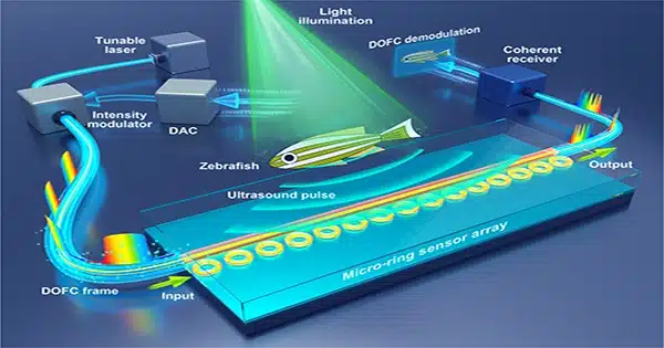
Researchers at the City University of Hong Kong have created a photoacoustic microscope with nearly 30 times the sensitivity of current equipment. This innovation enables high-resolution imaging of living animals while employing lower laser intensities, lowering the danger of tissue injury.
Photoacoustic microscopy uses low-power laser pulses to generate ultrasonic waves from light-absorbing molecules in tissue, which are subsequently translated into pictures. This approach allows for deeper subcellular resolution than standard optical microscopy, making it a viable tool for researching disorders such as cancer, diabetes, and stroke. However, its low sensitivity has been a major impediment to wider adoption.
To address this obstacle, the researchers modified their photoacoustic microscope with new hardware and a novel signal-processing method. They reconfigured the optical and acoustic parts to achieve accurate alignment and created a spectral-spatial filter algorithm to suppress noise and improve weak signals.

The novel technique combines coherent signals in both spectral and spatial dimensions, increasing the signal-to-noise ratio and enhancing the system’s total sensitivity 33-fold. This high-sensitivity device and algorithm allowed for thorough imaging of microvessels and oxygen saturation in live mice’s ears, eyes, and brains.
The researchers proved that their method, known as super-low-dose photoacoustic microscopy (SLD-PAM), yields high-quality images while consuming much less laser energy. The spectral-spatial filter revealed previously undetectable veins and arteries with an excitation energy of only 1 nJ. In contrast to the larger energy generally required for photoacoustic imaging, detailed pictures of the vascular network in the eyes and brains of mice were obtained with an excitation energy of only 4 nJ.
The researchers also demonstrated that SLD-PAM can achieve comparable image quality for molecular imaging utilizing weaker laser pulses, resulting in a greater dye survival rate and preventing photobleaching.
The researchers anticipate that SLD-PAM will revolutionize photoacoustic imaging by allowing noninvasive imaging while causing minimal damage to biological tissue. This approach has the potential to advance a wide range of biomedical applications while also opening up new paths for clinical translation.


