Each muscle is made of many long, cylindrical fibers arranged in parallel arrays. These fibers are composed of numerous fine fibrils, called myofibrils. Muscle fibers contract (shorten) in response to stimulation, then relax (lengthen) and return to their uncontracted state in a coordinated fashion. Their action moves the body to adjust to the changes in the environment and to maintain the positions of the various parts of the body. In general, muscles play an active role in all the movements of the body.
Muscles are of three types, skeletal, smooth, and cardiac.

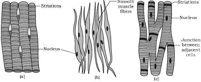
Fig: (a) Skeletal muscle, (b) smooth muscle, (c) Cardiac muscle
Voluntary or Striated Muscle or Skeletal muscle tissue is closely attached to skeletal bones. In a typical muscle such as the biceps, striated (striped) skeletal muscle fibers are bundled together in a parallel fashion (Figure: a). A sheath of tough connective tissue encloses several bundles of muscle fibers.
The smooth muscle fibers taper at both ends (fusiform) and do not show striations (Figure: b). Cell Junctions hold them together and they are bundled together in a connective tissue sheath. The wall of internal organs such as the blood vessels, stomach, and intestine contains this type of muscle tissue. Smooth muscles are ‘involuntary’ as their functioning cannot be directly controlled. We usually are not able to make it contract merely by thinking about it as we can do with skeletal muscles.
Cardiac muscle tissue is a contractile tissue present only in the heart. Cell Junctions fuse the plasma membranes of cardiac muscle cells and make them stick together (Figure: c). Communication Junctions (intercalated discs) at some fusion points allow the cells to contract as a unit, i.e., when one cell receives a signal to contract, its neighbors are also stimulated to contract.


