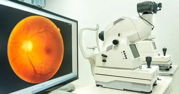
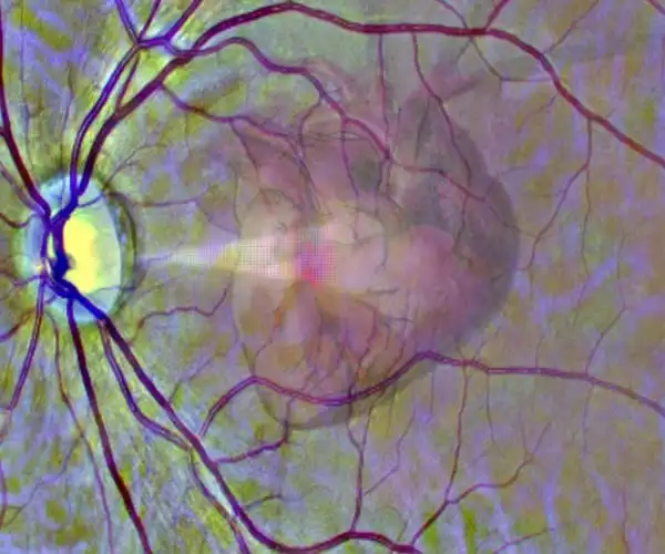
Scientists have created an artificial intelligence (AI) system that can analyze eye scans taken during a routine visit to an optician or eye clinic and identify patients who are at high risk of having a heart attack. Changes in the tiny blood vessels in the retina have been identified as indicators of broader vascular disease, including heart problems.
Deep learning techniques were used in the study, led by the University of Leeds, to train the AI system to automatically read retinal scans and identify those people who were likely to have a heart attack in the following year.
Deep learning is a complex set of algorithms that allows computers to recognize patterns in data and predict outcomes. The researchers report in the journal Nature Machine Intelligence that the AI system had an accuracy of between 70% and 80% and could be used as a secondary referral mechanism for in-depth cardiovascular investigation. The application of deep learning to the analysis of retinal scans has the potential to transform the way patients are routinely screened for signs of heart disease.
This technique has the potential to revolutionize cardiac disease screening. Retinal scans are relatively inexpensive and are commonly used in many optician practices. Patients who are at high risk of becoming ill as a result of automated screening may be referred to specialist cardiac services. The scans could also be used to detect early warning signs of heart disease.
Professor Alex Frangi
The study was overseen by Professor Alex Frangi, who holds the Diamond Jubilee Chair in Computational Medicine at the University of Leeds and is a Turing Fellow at the Alan Turing Institute. “Cardiovascular diseases, including heart attacks, are the leading cause of premature death worldwide and the second-leading cause of death in the UK,” he said. This causes chronic illness and misery all over the world.
“This technique has the potential to revolutionize cardiac disease screening. Retinal scans are relatively inexpensive and are commonly used in many optician practices. Patients who are at high risk of becoming ill as a result of automated screening may be referred to specialist cardiac services. The scans could also be used to detect early warning signs of heart disease.”
Scientists, engineers, and clinicians from the University of Leeds, Leeds Teaching Hospitals NHS Trust, the University of York, the Cixi Institute of Biomedical Imaging in Ningbo, part of the Chinese Academy of Sciences, the University of Cote d’Azur, France, the National Centre for Biotechnology Information and the National Eye Institute, both part of the National Institutes of Health in the United States, and KU Leuven in Belgium collaborated on the study. The data for the study came from the UK Biobank.
One of the study’s authors was Chris Gale, Professor of Cardiovascular Medicine at the University of Leeds and Consultant Cardiologist at Leeds Teaching Hospitals NHS Trust. “The AI system has the potential to identify individuals attending routine eye screening who are at higher future risk of cardiovascular disease, allowing preventative treatments to begin earlier to prevent premature cardiovascular disease,” he said.
Deep learning
During the deep learning process, the AI system examined over 5,000 people’s retinal and cardiac scans. The AI system discovered links between pathology in the retina and heart changes in the patient. Once the image patterns were learned, the AI system could estimate the size and pumping efficiency of the left ventricle, one of the four chambers of the heart, based solely on retinal scans. An enlarged ventricle is associated with a higher risk of heart disease.
The AI system could predict the patient’s risk of a heart attack over the next 12 months using information on the estimated size of the left ventricle and its pumping efficiency, as well as basic demographic data such as age and gender.
Currently, details about a patient’s left ventricle’s size and pumping efficiency can only be determined through diagnostic tests such as echocardiography or magnetic resonance imaging of the heart. These diagnostic tests can be costly and are frequently only available in a hospital setting, making them inaccessible to people in countries with less well-resourced healthcare systems – or increasing healthcare costs and waiting times in developed countries unnecessarily.
“The AI system is an excellent tool for unraveling the complex patterns that exist in nature, and that is what we have found here — the intricate pattern of changes in the retina linked to changes in the heart,” said Sven Plein, British Heart Foundation Professor of Cardiovascular Imaging at the University of Leeds and one of the study’s authors.