New CT scan technique reduces Radiation Exposure
A new CT (computed tomography) scan method can reduce radiation dose by splitting a full C-ray beam into thin and small beamlets. A CT scan technique that splits a full X-ray beam into thin beamlets can deliver the same quality of the image at a much-reduced radiation dose, according to a new UCL study. A team of researchers at the University College London (UCL) demonstrated that the new technique can provide the same quality image but with lower radiation doses.
A CT scan is a form of X-ray that creates very accurate cross-sectional views of the inside of the body. The technique, demonstrated on a small sample in a micro CT scanner, could potentially be adapted for medical scanners and used to reduce the number of radiation millions of people are exposed to each year. It is used to guide treatments and diagnose cancers and other diseases.
A computed tomography (CT) scan of the body uses sophisticated X-ray technology to help detect a wide range of diseases and conditions. It is used to guide treatments and diagnose cancers and other diseases. The method uses computers and rotating X-ray machines to generate cross-sectional images of the body.
Past studies have suggested CT scans may cause a small increase in lifelong cancer risk because their high-energy wavelengths can damage DNA. In the new study, published in Physical Review Applied, researchers placed a mask with tiny slits over an X-ray beam, breaking up the beam into beamlets. The procedure provides accurate images of the shoulders, head, spine, heart, abdomen, chest, and knee.
The researchers then compared the novel method to traditional CT scanning methods, wherein the item rotates as a full-beam is directed to it. The researchers compared the new technique to conventional CT scanning methods, where a sample rotates as a full-beam is directed on to it, finding it delivered the same quality of the image at a vastly reduced dose. It is one of the fastest and most reliable tools for examining many body parts, such as the pelvis, abdominal area, and chest since it provides cross-sectional views of all tissue types.
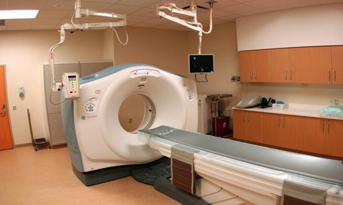
Fig: New CT scan technique may help lower radiation exposure
Dr. Charlotte Hagen (UCL Medical Physics & Biomedical Engineering), first author of the paper and a member of the UCL Advanced X-Ray Imaging Group, said: “Being able to reduce the dose of a CT scan is a long-sought goal. Our technique opens new possibilities for medical research and we believe that it can be adjusted for use in medical scanners, helping to reduce a key source of radiation for people in many countries.”
Conventional CT scans involve an X-ray beam is rotated around the patient. The new “cycloidal” method combines this rotation with a simultaneous backward and forwards motion. It can also provide detailed images to detect and diagnose vascular diseases, which can lead to complications if they are not treated immediately.
The use of beamlets enables a sharper image resolution, as part of the scanner “reading” the information from the X-ray is able to locate where the information is coming from more precisely. The imaging technique may also assess aortic aneurysms and pulmonary embolism, which are potentially fatal conditions that need urgent detection and treatment.
Professor Sandro Olivo (UCL Medical Physics & Biomedical Engineering), the senior author of the paper, said: “This new method fixes two problems. It can be used to reduce the dose, but if deployed at the same dose it can increase the resolution of the image. They found that the new method that delivers lesser doses of radiation produced the same quality of the image.