Researchers determine which neurons dilated the cerebral blood vessels
The brain, an organ of unprecedented complexity, seems to have a basic design glitch: it absorbs a huge amount of energy but lacks a fuel reserve for usage when required. Neurons regulate blood pressure in tiny channels in the brain, but we know very little about this relationship. Now, a team of Penn State engineers has discovered a correlation between nitric oxide expressing neurons and improvements in arterial diameters in mice that could shed light on brain function and aging.
Some Neurons Target Tiny Cerebral Blood Vessel Dilation/ The researchers looked for a way to turn neurons off and on so that they could determine which neurons dilated the blood vessels.
The brain is one of the most heavily perfumed organs in the body, and proper brain operation demands that brain blood supply be sustained at a steady pace, independent of the metabolic needs of other organs. “There are many types of neurons in the brain,” said Patrick J. Drew, Huck Distinguished Associate Professor of Engineering Science and Mechanics, Neurosurgery and Biomedical Engineering. “People have seen that blood flow and neural activity go together, and we wanted to measure blood flow and activity in specific subtypes of neurons.”
Neuronal activation within the brain evokes a local rise in blood pressure, a reaction called functional hyperemia. This reaction guarantees that the activated neurons receive adequate oxygen and nutrients to sustain the function and health of the tissues. Researchers were searching for a way to turn the neurons off and on so that they could determine which neurons dilated the blood vessels. “We knew that when animals run, all neurons become more active, and this causes the blood vessels to react,” Drew said. “There are excitatory and inhibitory nerves, and there is some evidence that vasodilation happens as you activate inhibitory neurons, but there are a lot of inhibitory neurons. We were trying to figure out which ones were responsible.”
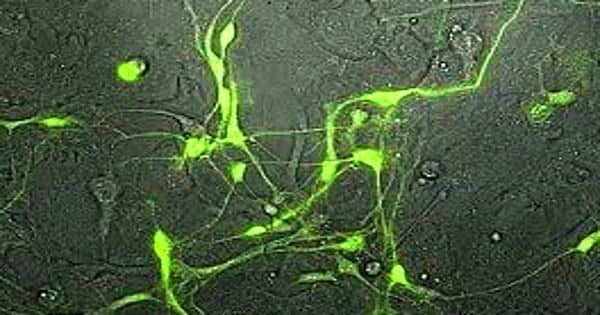
The researchers used two-photon microscopy to look at the surface and deep arteries of the somatosensory cortex of mice when the mice were awake. They were able to make several comparisons of the same arteries in the same animals over a series of separate neuronal disruptions so that they could analyze findings in individuals rather than through individuals. They posted their findings to eLife.
“Thirty years ago, people realized that nitric oxide was a vasodilator,” Drew said. “Other researchers have seen a nitric oxide enzyme stimulate a subset of neurons, suggesting that this might help to locate the responsible neurons.”
Drew and his colleagues have found that there has been a transition in dilatation with a rise or decrease in the nitric oxide enzyme without a change in electrical activity. According to Drew, the brain is oversupplied with oxygen, so increasing dilation when running or for other purposes does not actually increase the supply of oxygen.
“The changes do not seem to be related to the need,” Drew said. “It’s a mystery why this is going to happen.”
Since researchers have been able to observe variations in the same blood vessel over time and with various stimuli, they could equate these changes to the baseline in the individual animals. Some classes of neurons may also be blocked for varying durations of time.
“Our results suggest a model in which approximately half of the dynamic range in the basal and evoked blood arterial diameter is controlled by a small group of neurons and the rest is controlled by other neurons and astrocytes,” the researchers write. “Any damage or dysfunction of the nNOS neurons (producing nitric oxide enzymes) could result in decreased basal blood flow, regardless of metabolic need.”
The researchers have observed that variations in the diameter of the blood vessels influence the amplitude of the fmri signals. Since fmri is used to image blood vessels in animals and humans, it can generate signals that are not linked to neuronal activity. “This tells us that the fmri signals may not represent the overall activity,” Drew said. “It suggests that if these neurons die, it could cause dementia to develop.”
The brain is highly dependent on the constant flow of blood to function. The cerebral vasculature is thus endowed with neurovascular regulation mechanisms that ensure that the blood supply of the brain is commensurate with the energy requirements of its cellular constituents. Researchers would like to know what happens if these nerves have been shut down for a long time. They would still like to see what happens if the same neurons are activated for a long time. Another aim is to explain what drives these neurons, what modular feedback do they get, and in what other areas of the brain do they function?