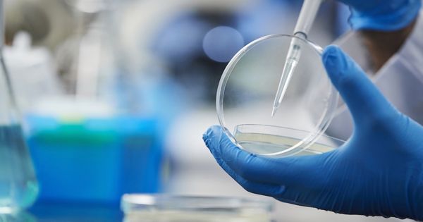
A team has created a handheld computer that generates high definition 3D images of human skin within 10 minutes. A team from Nanyang Technology University, Singapore (NTU Singapore) has created a portable computer that generates high-resolution 3D images of human skin within 10 minutes. The team suggested that a handheld skin imaging (imagery) system could be used to determine the seriousness of skin disorders, such as eczema and psoriasis.
When anyone has a skin disorder such as eczema or psoriasis, it helps to be able to see all the slight ridges and grooves in the infected region. This is where a compact new technology comes in, built to create 3D images of the skin in a matter of minutes.
A portable device will help improve the diagnosis, treatment of skin conditions and injuries. NTU Singapore team develops a portable device that creates 3D images of skin in 10 minutes.
3D skin imaging may be useful for doctors, as much of the technology used to diagnose skin conditions only offers 2D representations of the skin surface. As the unit also charts the depth of the ridges and grooves of the skin up to 2 mm, it can also help to monitor wound healing.
Built at Singapore’s Nanyang Technology University, the low-cost device battery-powered prototype weighs only 100 grams (3.5 oz) and measures 7 by 10 cm (2.8 by 3.9 in). On the other hand, much larger OCT (optical coherence tomography) devices – currently used for 3D skin imaging – cost thousands of dollars and can weigh up to 30 kilograms (66 lb).
The unit presses a specially made film onto the skin of the subject to achieve an imprint of up to 5 by 5 centimeters, which is then exposed to an electrical charge, producing a 3D image. Researchers have developed and 3D printed a prototype of their system using polylactic acid (PLA), a biodegradable bioplastic. The battery-operated unit, measuring 7cm by 10cm, weighs just 100 grams.
NTU Singapore Team Develops Portable Device That Creates 3D Images of Skin in 10 Minutes
The made-in-NTU prototype is produced at a fraction of the cost of devices with similar technology, such as optical accuracy tomography (OCT) machines, which can cost thousands of dollars and weigh up to 30 kilograms.
Assistant Professor Grzegorz Lisak of the NTU School of Civil and Environmental Engineering, who led the study, said: “Our non-invasive, simple and affordable technology could be used to supplement existing methods of diagnosing and treating skin diseases. In remote areas that do not have ready access to health care, non-medical personnel will create skin maps using the device and send them to the hospital.
Dr. Yew Yik Weng, Consulting Dermatologist at the National Skin Center, and an Assistant Professor at NTU’s Lee Kong Chian School of Medicine said: “Technology is a fascinating way to map the surface texture of human skin. It may be a valuable tool for mapping skin texture and wound healing in a 3D way, which is an interesting way to map the surface texture of human skin.
Asst. Prof. Dr. Yew added: “The device could be particularly useful in studies involving wound healing, as we currently lack a tool that maps the length and depth of the skin ridges. At present, we rely on photographs or measurements in our studies that could only provide a 2D assessment.” The first author of the research, Mr. Fu Xiaoxu, a Ph.D. student at the NTU School of Civil and Environmental Engineering.
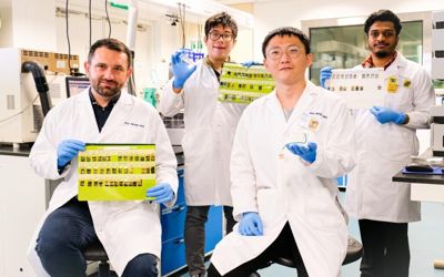
Published in the scientific journal Analytica Chimica Acta, the technology was established by Asst Prof Lisak, who is also Director of the Residues & Resource Reclamation Center at the Nanyang Environmental and Water Research Institute (NEWRI), and his Ph.D. student, Mr. Fu Xiaoxu.
The ‘golden’ solution to 3D skin mapping
The main component of the NTU system is a polymer called PEDOT:PSS, widely used in solar panels to transform light to electricity. However, the team found a different application for its electrical conductivity—to replicate skin patterns on gold-plated film. Gold is used because it has exceptional electrical conductivity and flexibility.
To use the device, a person presses a button to push the gold-plated film onto the skin of the subject to acquire an imprint. This allows sebum, an oily material formed by the skin, to be transported to the film, producing an image of the surface of the skin.
The imprint of the skin is then moved to a handheld unit where a group of electrodes is submerged in a solution. Through a further press of a button, the system activates an electrical charge flow, which allows PEDOT:PSS to be collected on the surfaces of the gold-plated film in places not protected by sebum. This results in a high-resolution 3D skin map reflecting the ridges and grooves of the skin.
Using pig skin as a guide, the researchers have shown that the technology has been able to trace the pattern of different wounds, such as punctures, lacerations, abrasions, and incisions. The team also revealed that even the intricate web of wrinkles on the back of a human hand could be caught on the video. The thin film is also versatile enough to map features on irregular parts of the skin, such as elbow creases and fingerprints.
Asst Prof Lisak added: “The device has also proved to be successful in extracting fingerprints and provides a high-resolution 3D image of their characteristics.” Commenting on the possible applications of the device, Asst Prof Dr. Yew added: “The device can aid in the detection of fingerprints, which is typically done in forensic research.
To date, it has been successfully used on pig skin to map wounds such as punctures, lacerations, abrasions, and incisions. It has also been used to picture the skin on the back of the human hand, but the film is durable enough that it can also be extended to more uneven areas such as the elbows. In order to further confirm its effectiveness, the team is exploring clinical trials later this year to assess the viability of their unit, as well as other possible therapeutic applications.