The technique of electron microscopy (EM) is used to obtain high-resolution images of biological and non-biological specimens. In biomedical research, it is used to investigate the detailed structure of tissues, cells, organelles, and macromolecular complexes. The use of electrons (which have very short wavelengths) as the source of illuminating radiation results in the high resolution of EM images. To answer specific questions, electron microscopy is used in conjunction with a variety of ancillary techniques (e.g., thin sectioning, immuno-labeling, negative staining). EM images provide critical information about the structural basis of cell function and disease.
Physicists at Friedrich-Alexander-Universität Erlangen-Nürnberg (FAU) have developed a framework that allows scientists to use a traditional scanning electron microscope to observe interactions between light and electrons. The procedure is significantly less expensive than previous technologies, and it also allows for a broader range of experiments. The findings have been published in the journal Physical Review Letters.
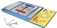
We created a special spectrometer based on magnetic forces that is integrated directly into the microscope. The underlying principle is that the magnetic field diverts electrons to varying degrees depending on their speed.
Prof. Dr. Peter Hommelhoff’s
The quantum computer is just one example of how important understanding the fundamental processes underlying interactions between photons and electrons is. When combined with ultra-short laser pulses, it is possible to measure how photons change the energy and speed of electrons. Until recently, photon-induced electron microscopy (PINEM) relied solely on transmission electron microscopes (TEM). Although they have the resolution to pinpoint individual atoms, they are significantly more expensive than scanning electron microscopes (SEM), and their sample chamber is extremely small, only a few cubic millimeters in size.
Measuring differences down to a only a few hundred thousandths of a whole
Researchers at Prof. Dr. Peter Hommelhoff’s Chair of Laser Physics have now modified a traditional SEM to conduct PINEM experiments. They created a special spectrometer based on magnetic forces that is integrated directly into the microscope. The underlying principle is that the magnetic field diverts electrons to varying degrees depending on their speed.
An accurate reading of this deviation is provided using a detector that converts electron collisions into light. The method enables researchers to measure even the smallest changes in energy, down to differences of a few hundred thousandths of the original value – enough to distinguish the contribution of a single light energy quanta – a photon.
A wider spectrum of experiments possible in the future
In more ways than one, the Erlangen physicists’ discovery is groundbreaking. Being able to study photon-electron interactions without using TEM, which costs several million euros, could make research more affordable. Furthermore, because the chamber of a SEM has a volume of up to 20 cubic centimeters, a much broader range of experiments can now be performed, as additional optical and electronic components such as lenses, prisms, and mirrors can be placed directly next to the samples. The researchers anticipate that the entire field of microscopic quantum experiments will shift from TEM to SEM in the coming years.
Conventional scanning electron microscopy is based on the emission of secondary electrons from a specimen’s surface. A scanning electron microscope is the EM equivalent of a stereo light microscope due to its high depth of focus. It can produce detailed images of the surfaces of cells and whole organisms that TEM cannot. It can also be used for particle counting, determining particle size, and controlling processes. The image is formed by scanning a focused electron beam onto the surface of the specimen in a raster pattern, hence the name scanning electron microscope.
The interaction of the primary electron beam with the atoms near the surface causes particle emission at each point in the raster (e.g., low energy secondary electrons, high energy back scatter electrons, X-rays and even photons). These can be collected using a variety of detectors and their relative number translated to brightness at each equivalent point on a cathode ray tube. Because the raster at the specimen is much smaller than the viewing screen of the CRT, the final image is a magnified image of the specimen. Appropriately equipped SEMs (with secondary, backscatter, and X-ray detectors) can be used to study the topography and atomic composition of specimens, as well as the surface distribution of immuno-labels, for example.