The cardiac sympathetic neural network, which controls the body’s heart rate and ‘fight-or-flight’ response, has been digitally topographically mapped by researchers. They hope that this map will one day serve as a guide for treating cardiovascular diseases with bioelectronic devices.
The cardiac sympathetic neural network, which controls the body’s heart rate and “fight-or-flight” response, has been digitally topographically mapped by a team of UCF College of Medicine researchers. They hope that this map will one day serve as a guide for treating cardiovascular diseases with bioelectronic devices.
The study, led by Dr. Zixi Jack Cheng, a neuro-cardiovascular scientist, was published in the Scientific Reports journal and was the project of an interdisciplinary team of researchers from UCF along with several other institutions as well as industry partners MBF Bioscience and SPARC Data and Resource Center. The project has been supported by the NIH Common Fund’s Stimulating Peripheral Activity to Relieve Conditions (SPARC) program, and funding from National Institute on Neural Disorders and Stroke and National Heart, Lung and Blood Institute.
“This mapping goes above and beyond what you can find in a textbook,” Dr. Cheng explained. “This is an interactive digitized brain-heart atlas.” We hope it will be useful not only for scientists and physicians, but also for students learning about heart neuroanatomy.”
The cardiac-sympathetic nerve system is very complex and remains poorly understood. So having this detailed mapping in the heart could give us important insights into the architecture of the cardiac-sympathetic nerve and provide the foundation for future functional and molecular studies of sympathetic control of the heart.
Dr. Jin Chen
The map could be used to guide treatments like neuromodulation therapy, which involves electrically stimulating nerves to treat cardiovascular conditions.
Dr. Cheng’s team and his SPARC collaborators previously created a detailed 3D map of the intrinsic nervous system of a rodent heart. The system, dubbed the “little heart brain,” is made up of thousands of neurons that surround the heart and regulate heartbeat and blood circulation. Their most recent project expanded on that research by mapping the topographical network of nerves in the sympathetic nervous system and their connection to the heart. The researchers hope that the advanced blueprint will aid scientists and physicians in studying the brain-heart connection and navigating more precise control of various heart regions, including those that control the heartbeat.
Through an intricate network of nerves, the sympathetic nervous system regulates cardiac functions. It can help the body respond to dangerous or stressful situations by increasing heart rate and delivering more blood to areas that require more oxygen. The system is also in charge of heart rate, blood pressure, digestion, and other vital functions.
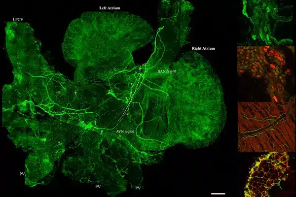
The team used a combination of cutting-edge techniques to image, trace, digitize, and quantitatively map the distribution of the sympathetic nervous system, which included the entire atria and ventricles of the heart.
“The groundbreaking aspect of this project is the precision with which the mapping is completed at the microscopic level, allowing us to see single cells and single nerve axons,” Dr. Cheng said. This is the first time scientists have seen the entire organ at such a detailed level.”
Dr. Yuanyuan Zhang, a postdoctoral fellow in Dr. Cheng’s lab, explained that this map will allow researchers to test the functional role of a specific nerve by activating or deactivating it and observing the effect on the body. According to him, the mapping can also be used as a guide for treatments like neuromodulation therapy, which involves electronically stimulating nerves to treat cardiovascular conditions like hypertension, sleep apnea, and heart failure.
“Utilizing our map as a sympathetic-cardiac atlas opens the door to innovative therapies for several cardiovascular diseases, and nerve-related disorders, and avoids side effects associated with many pharmaceuticals,” said Ariege Bizanti, a Ph.D. candidate in Dr. Cheng’s lab.
According to CDC statistics, heart disease is the leading cause of death for both men and women in the United States, accounting for 697,000 deaths in 2020. In the United States, one person dies from cardiovascular disease every 34 seconds.
“The cardiac-sympathetic nerve system is very complex and remains poorly understood,” said Dr. Jin Chen, another UCF project collaborator. “So having this detailed mapping in the heart could give us important insights into the architecture of the cardiac-sympathetic nerve and provide the foundation for future functional and molecular studies of sympathetic control of the heart.”