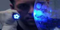
Reactive molecules, such as free radicals, can be formed in the body after exposure to certain conditions or compounds which can cause cell damage. Antioxidants can mitigate the damage by intervening with the radicals until they affect the cells.
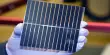
Driven by Enrique Gomez, Professor of Chemical Engineering and Materials Science and Engineering, Penn State researchers applied this principle to avoid imaging harm to the conduct of polymers comprising soft electronic devices such as organic solar cells, organic transistors, bioelectronic devices and flexible electronics.
Polymers can only be imaged to a certain point with high-resolution transmission electron microscopy (HRTEM) because the bombardment of electrons used to form images breaks the sample apart.
According to Gomez, the simulation of polymer-conducting structures are important for the further creation of these materials and for the commercialization of soft electronic devices-but real imaging can cause harm that restricts what researchers can see and understand.
“It turns out antioxidants, like those you’d find in berries, aren’t just good for you but are also good for polymer microscopy,” Gomez said. Polymers can only be imaged at a certain stage with high-resolution transmission electron microscopy (HRTEM) and the electron bombardment used to form images of tears in the sample apart.
The researchers investigated the damage with a view to determining its underlying cause. They observed that the HRTEM electron beam created free radicals that degraded the molecular structure of the sample. The application of butylated hydroxytoluene, an antioxidant frequently used as a food additive, to the polymer sample avoided this damage and eliminated a further constraint on imaging conditions—temperature.
“Until now, the main strategy for minimizing polymer damage has been imaging at very low temperatures” said paper co-author Brooke Kuei, who earned her doctorate in materials science and engineering in the Penn State College of Earth and Mineral Sciences in August. “Our work demonstrates that the beam damage can be minimized with the addition of antioxidants at room temperature.”
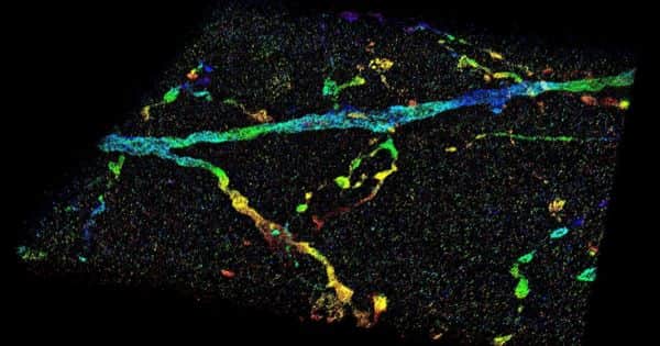
Although the researchers did not quantitatively measure the resolution limits arising from this process, they were able to image the polymer at a resolution of 3.6 angstroms, an increase from their previous resolution of 16 angstroms. For instance, an angstrom is around one millionth the breadth of human hair.
Polymers are made up of molecular chains on top of each other. Previously, the distance of 16 angstroms was the distance between the chains, but visualization at 3.6 angstroms enabled researchers to envision patterns of near contacts along the chains. For the electrically conductive polymer studied in this analysis, researchers could trace the path in which electrons move. According to Gomez, this helps them to better understand the conductive structures of polymers.
“The key to this advancement in polymer microscopy was understanding the fundamentals of how the damage occurs in these polymers,” Gomez said. “This technological advance will hopefully help lead to the next generation of organic polymers.” National Research Foundation and Kuei Graduate Fellowship via the U.S. The Department of Energy Science Office has funded this work.